OCT
Optical coherence tomography (OCT) is a non-invasive imaging test. OCT uses light waves to take cross-section images of your retina.
With OCT, your ophthalmologist can see each of the retina’s distinctive layers. This allows your ophthalmologist to map and measure their thickness. These measurements help with diagnosis. They also provide treatment guidance for glaucoma and diseases of the retina. These retinal diseases include age-related macular degeneration (AMD) and diabetic eye disease. As well, OCT provides images of the cornea and angle.
ZEISS CIRRUS 5000: https://www.zeiss.com/meditec/int/product-portfolio/optical-coherence-tomography-devices.html
A-Scan
A-scan is the short form for amplitude scan. This eye ultrasound gives details about the length of the eye. It is a one-dimensional scan of the eye. The measurement of the axial length of the eye through an A-scan is necessary for placing with high lever accuracy of the intraocular lens (IOL, artificial lens) implanted during a cataract surgery. Our latest software upgrade provides different IOL formulas for IOL calculations.
ZEISS IOLMaster 700: https://www.zeiss.com/meditec/int/product-portfolio/optical-biometers/iolmaster-700.html
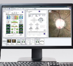
Visual Fields
Your visual field is how wide of an area your eye can see when you focus on a central point. Visual field testing is one way your ophthalmologist measures how much vision you have in either eye, or how much peripheral vision loss may have occurred over time. The new technology of our HFA3 increases the accuracy of the field test in patients of all ages.
ZEISS Humphrey® Field Analyzer 3 (HFA3): https://www.zeiss.com/meditec/us/product-portfolio/perimetry/humphrey-visual-field-analyzer-3-with-sita-faster.html
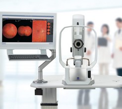
Retinal Imaging
A non-invasive diagnostic tool used to take high-definition scanning images of the back of your eye. The technology used includes specialized cameras and scanners to magnify your retina, optic nerve, and blood vessels inside the eye with real color.
ZEISS CLARUS 500: https://www.zeiss.com/meditec/int/product-portfolio/retinal-cameras/clarus-500.html
Corneal Topography
Corneal topography is a special photography technique that maps the surface of the clear, front window of the eye (the cornea). It works much like a 3D (three-dimensional) map of the world, that helps identify features like mountains and valleys.
ZEISS ATLAS 9000: https://www.zeiss.com/content/dam/Meditec/us/brochures/atlas.pdf
FORUM®
FORUM® from ZEISS is a scalable and flexible ophthalmic data management solution. It streamlines practice workflows by connecting ZEISS devices and providing access to all patient examination data which allows the physician to make confident decisions at a glance. We are the only Ophthalmological practice in Central Alberta with this data management solution that includes new and advanced software for Glaucoma.
https://www.zeiss.com/meditec/int/product-portfolio/data-management-software/forum.html
B-Scan
B-Scan or brightness scan is another method used for ocular assessment via ultrasound. It can be performed directly on the anesthetized eye. In cases of trauma or in children, B-scan can be performed over the eyelid with coupling jelly. Measurements derived from B-scan include visualization of the lesion, including anatomic location, shape, borders, and size. It can be used for a detection of a wide range of pathological structures, including retinal or choroidal detachment, foreign bodies, calcium, and tumors.
https://www.accutome.com/b-scan-plus
YAG Laser
Posterior capsulotomy is an in-office procedure that you might need sometime after cataract surgery. It helps you see clearly if your vision becomes cloudy again.
When you have cataract surgery, your ophthalmologist removes your eye's cloudy lens. They replace it with a clear, artificial intraocular lens (IOL). The IOL is held in place in the eye’s natural lens capsule. Weeks, months or years later, this capsule can become cloudy or wrinkled, causing blurry vision. This is called a posterior capsule opacification (PCO). It’s also sometimes called a "secondary cataract" or "scar tissue." With posterior capsulotomy, a laser is used to make an opening in the cloudy capsule. This allows light to pass through again for clear vision. It is also used to perform Laser Iridotomy for prevention and treatment of acute angle glaucoma.
https://www.clarionmedical.com/en-CA/Vision/Lasers/Lumenis-Selecta-Duet
SLT Laser
Selective laser trabeculoplasty (SLT) is an in-office procedure that reduces intraocular pressure in patients with glaucoma. The laser is applied through a special contact lens to the drainage system of the eye where it stimulates a biochemical change that improves the outflow of fluid from the eye.
https://www.clarionmedical.com/en-CA/Vision/Lasers/Lumenis-Selecta-Duet